Computed Tomography (CT) Angiogram
A computed tomography angiogram (CT angiogram) is a test that uses X-rays to make detailed pictures of the heart and the blood vessels that go to the heart, lungs, brain, kidneys, head, neck, legs, and arms.
Q & A
Test Overview
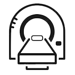
A computed tomography angiogram (CT angiogram) is a test that uses X-rays to make detailed pictures of the heart and the blood vessels that go to the heart, lungs, brain, kidneys, head, neck, legs, and arms. This test can show where a blood vessel is too narrow or blocked. It can also show if a blood vessel has a bulge (called an aneurysm) or a buildup of fat called plaque. During a CT angiogram, you lie on a table that moves through an opening in the scanner that looks like a doughnut. A special dye called a contrast agent is put into a vein (IV) in your arm or hand to make it easier to see the blood vessels on the scan. If you are getting this test to look at your heart and the blood vessels that lead to it (coronary arteries), you may be given a beta-blocker to slow your heart rate during the test.
What are the pros and cons?
A standard angiogram is more invasive than a CT angiogram. In a typical angiogram, a thin tube called a catheter is put through an artery in your arm or leg and up to the area being looked at. In a CT angiogram, on the other hand, no tubes are put into your body. If your doctor sees that one or more of your blood vessels are narrowed or blocked, you may still need a standard angiogram to double-check the abnormal results from the CT angiogram. This is more likely to happen if your doctor is thinking about operating to fix the blockage or narrowing.
During a CT angiogram, if your doctor finds a big blockage in one of your blood vessels, you won’t be able to get an angioplasty right away to get rid of the blockage. You will need a different plan. But if you have a standard angiogram and the doctor finds a major blockage, he or she can do an angioplasty during the angiogram.
Why It Is Done
A blood clot in the lungs (pulmonary embolism).
A blockage or narrowing of one of the coronary arteries. This can happen when fat (cholesterol) and calcium build up in the arteries. Plaque is the name for this buildup.
Heart problems, like pericarditis, which is an inflammation of the sac that surrounds the heart, and damaged or broken heart valves.
The aorta, a large blood vessel that carries blood from the heart to the rest of the body, has a bulge (aneurysm) or a tear (dissection).
During the test
Someone will put a contrast dye in a vein in your arm or hand. If you are getting a CT angiogram to look at your heart and the blood vessels that lead to it (coronary arteries), you may be given a beta-blocker to slow your heart rate during the test. The table slides into the scanner’s round hole. During the scan, the table will move. The scanner moves around your body inside the doughnut-shaped case.
During the scan, you will be asked to stay still. You might have to hold your breath for short amounts of time. You might be the only one in the room for scanning. But during the test, a technologist will watch you through a window and talk to you.


